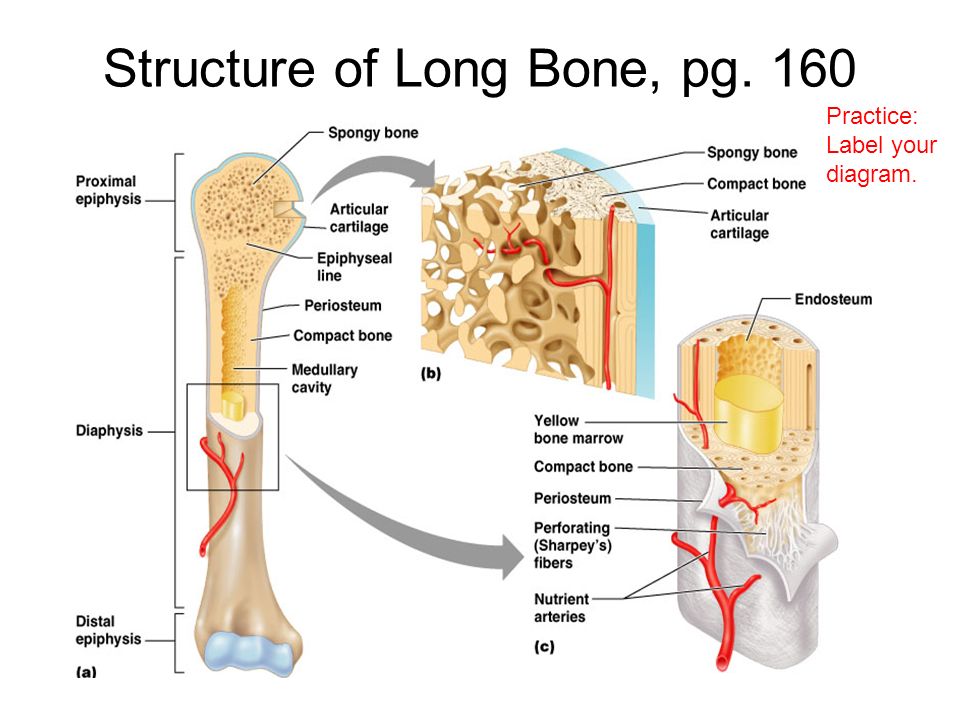
Long Bone With Diagram / Label number 3 in the diagram is pointing to :. Parts of a long bone. This is an online quiz called long bone anatomy. The diaphysis and the epiphysis (figure 6.3.1). Inside this is a layer of spongy (cancellous) bone which contains red bone marrow. Firstly, divide the skeleton into small groups―the skull, the chest and back, the hands, the pelvis, and lastly, the legs.
The long bones are those that are longer than they are wide. Learn long bone diagram with free interactive flashcards. It can be found under the periosteum and in the diaphyses of long bones, where it provides support and protection. The shiny, articulating cartilage on the ends of a bone. Long bone diagram unlabled manual e books.
Chap 6 Bones Skeletal Tissue Learning Objectives 1 Compare Contrast The Structure Of The 4 Bone Classes And Provide Examples Of Each Class 2 Explain Ppt Download from images.slideplayer.com Click on the tags below to find other quizzes on the same subject. The diaphysis and the epiphysis (figure 6.3.1). Long bones grow more than the other classes of bone throughout childhood and so are responsible for the bulk of our height as adults. You need to get 100% to score the 10 points available. Label number 3 in the diagram is pointing to : We cover the diaphysis, the epiphysis, spongy and c. Hollow bone or long bone is longer than it is wide and is composed of the following elements image: The diaphysis and the epiphysis.
The bone on the left in the image is the :
There is a printable worksheet available for download here so you can take the quiz with pen and paper. The bone on the left in the image is the : The structure of a long bone allows for the best visualization of all of the parts of a bone (figure 1). Long bone diagram unlabled manual e books. A typical long bone shows the gross anatomical characteristics of bone. You need to get 100% to score the 10 points available. Long bones are one of the five bone types that are classified by shape. Describe the timing and causes of epiphyseal plate closure. Smooth, white tissue that covers the ends of bones where they come together to form joints. Typical long bone labeled long bone anatomy human. Compact bone is the denser, stronger of the two types of bone tissue ( link ). Students fill in the boxes with the names of the bones. Found in the ends of long bones;
Examples of long bones include the femur, tibia, fibula, metatarsals, and phalanges. There is a printable worksheet available for download here so you can take the quiz with pen and paper. Related posts of diagram of of a long bone bone on side of the foot. Typical long bone labeled long bone anatomy human. Long bone diagram epiphyseal plate :
1 from This is an online quiz called label the long bone. Long bone diagram unlabled manual e books. Long bones are one of the five bone types that are classified by shape. Inside this is a layer of spongy (cancellous) bone which contains red bone marrow. Study long bone diagram flashcards from alan lin's umass amherst class online, or in brainscape's iphone or android app. Grasping organ at the end of the forelimb of certain vertebrates that exhibits great mobility and flexibility in the digits and in the whole organ. A long bone has diaphyseal bone is organized to create the best balance between weight and structural strength. A long bone has two parts:
Helps keep bones light in weight.
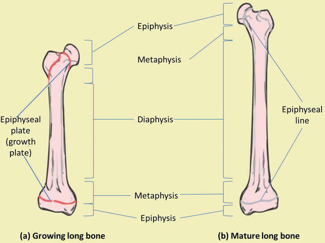
They are numbered from one to five, starting from the medial (inner) side of the foot. The long bones are those that are longer than they are wide. Describe the timing and causes of epiphyseal plate closure. Bone · august 12, 2020. In long bones, as you move from the outer cortical compact bone to the inner medullary cavity, the bone transitions to spongy bone. At level 30 construction, he will give a lecture about the skill granting 4,500 construction experience per bone, equivalent to 32,175 coins worth. The end of a long bone. Endosteum is a thin, soft, connective tissue, lining the cavity of long bones like humerus and femur. What is label number 4 pointing to in the diagram? Compact bone is the denser, stronger of the two types of bone tissue ( link ). Long bones have a thick outside layer of compact bone and an inner medullary cavity containing bone marrow. Bone is specialised a type of connective tissue. It can be found under the periosteum and in the diaphyses of long bones, where it provides support and protection.
Bone is specialised a type of connective tissue. Smooth, white tissue that covers the ends of bones where they come together to form joints. Bones of foot labeled diagram poster. Choose from 500 different sets of long bone diagram flashcards on quizlet. Students fill in the boxes with the names of the bones.
Bone Development And Growth Intechopen from www.intechopen.com The blood vessels inside a bone. Hollow bone or long bone is longer than it is wide and is composed of the following elements image: Study long bone diagram flashcards from alan lin's umass amherst class online, or in brainscape's iphone or android app. There is a printable worksheet available for download here so you can take the quiz with pen and paper. Grasping organ at the end of the forelimb of certain vertebrates that exhibits great mobility and flexibility in the digits and in the whole organ. A long bone has two parts: The end of a long bone. Found in the ends of long bones;
The end of a long bone.
Found in the ends of long bones; A long bone is a bone that is significantly longer than it is wide. Compact bone is the denser, stronger of the two types of bone tissue ( link ). The first metatarsal bone, the shortest, thickest and strongest metatarsal, links to the big toe. Students fill in the boxes with the names of the bones. Bone on side of the foot 12 photos of the bone on side of the foot bone on side of foot growing, bone on side of foot sticks out, fractured bone on side of foot, the bone on the side of my foot is sticking out, what is the bone on the side of my … Characterized by irregular spaces filled with red bone marrow that makes blood cells; This long bone connects with the knee at one end and the ankle at the other. The bone on the right in the image is the : A long bone has a shaft and 2 ends. Bones of foot labeled diagram poster. Smooth, white tissue that covers the ends of bones where they come together to form joints. In long bones, as you move from the outer cortical compact bone to the inner medullary cavity, the bone transitions to spongy bone.


0 Komentar